Neck Muscle Diagram : 38 174 Best Neck Muscles Images Stock Photos Vectors Adobe Stock. Accidents can lead to neck pain, as can poor posture and arthritis. Superficial muscles are the muscles closest to the skin surface and can usually be seen while a body is performing actions. Nerves in the neck, medically referred to as the cervical spine, help transmit information along the pathways of the central and peripheral nervous system, including sensory and motor skills processes.the cervical spine consists of eight different sets of nerves. A neck strain or sprain occurs when one or more neck muscles, ligaments or tendons are injured. The anatomy of the neck and shoulders is very interesting.
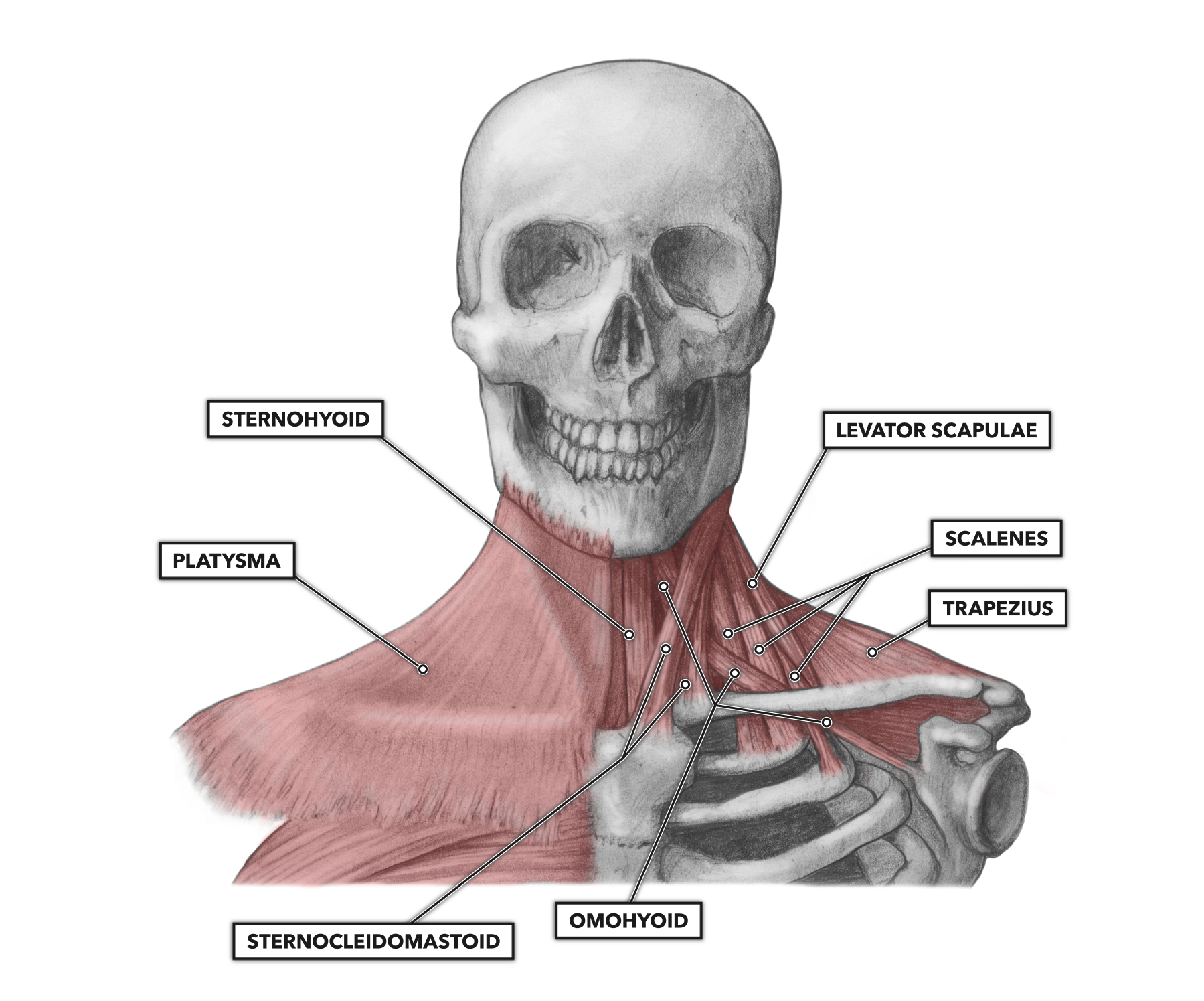
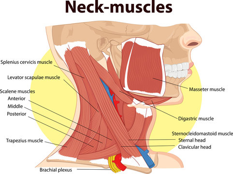
A neck strain or sprain occurs when one or more neck muscles, ligaments or tendons are injured. The sternocleidomastoid muscle, or scm, is one of the larger and more superficial muscles in the neck, making it an important and easily identifiable anatomical landmark.it arises from two origins: There are many muscles around the neck that help to support the cervical spine and allow you to move your head in different directions. Muscle head anatomy vocal organ diagram female neck anatomy neck wireframe head neck human anatomy head artery anatomy face pharynx vector neck degree head anatomy 3d. Anterior, lateral and posterior groups, based on their position in the neck.the musculature of the neck is further divided into more specific groups based.
Primary Neck Cancer Anatomy from 2pybk2la9r-flywheel.netdna-ssl.com Muscle identification, muscle actions, and muscle origins and insertions. Muscular back of a man muscular back of a man for study, great to be used in medicine works and health. Head and neck muscles diagram in this image, you will find cranial aponeurosis, temporalis, occipitalis, masseter, sternocleidomastoid, trapezius, platysma, orbicularis oris, buccinator, zygomaticus, orbicularis oculi, frontalis in head and neck muscles diagram. Muscles of the neck (musculi cervicales) the muscles of the neck are muscles that cover the area of the neck hese muscles are mainly responsible for the movement of the head in all directions they consist of 3 main groups of muscles: Related posts of diagram of the neck anatomy veins and arteries of the neck. Neck muscles are bodies of tissue that produce motion in the neck when stimulated. Learn vocabulary, terms, and more with flashcards, games, and other study tools. Anatomy of neck and shoulder stock illustrations.
The anatomy of the neck and shoulders is very interesting.
Most minor neck strains heal in a relatively short amount of time. Neck muscles are bodies of tissue that produce motion in the neck when stimulated. Search for anatomy of the head and neck in these categories. Deep in perspective to the trapezius muscle, the. Neck muscles help support the cervical spine and contribute to movements of the head, neck, upper back, and shoulders. Our third youtube film is ready to run. 1) above the cervical area (longissimus capitis), 2) in the cervical area (longissimus cervicis), and 3) in the upper back or thoracic area (longissimus thoracis). Neck and shoulder muscles diagram the superficial back muscles attachments actions teachmeanatomy Related posts of arteries in the neck picture veins and arteries of the neck. The neck muscles, including the sternocleidomastoid and the trapezius, are responsible for the gross motor movement in the muscular system of the head and neck. Head and neck muscles diagram in this image, you will find cranial aponeurosis, temporalis, occipitalis, masseter, sternocleidomastoid, trapezius, platysma, orbicularis oris, buccinator, zygomaticus, orbicularis oculi, frontalis in head and neck muscles diagram. Watch the whole lecture (all 8 videos) by goin. Learn vocabulary, terms, and more with flashcards, games, and other study tools.
Anterior, lateral and posterior groups, based on their position in the neck.the musculature of the neck is further divided into more specific groups based. The levator scapulae muscle is attached at the top four cervical vertebrae (c1 to c4) and runs down the side of the neck to attach at the top of. The rotation function takes the head into the opposite side to which this neck and shoulder muscle is located. Neck and shoulder muscles diagram the superficial back muscles attachments actions teachmeanatomy Part 7 in an 8 part lecture on skeletal muscle in a flipped human anatomy course taught by wendy riggs.
Crossfit Cervical Muscles Part 1 from www.crossfit.com Superficial muscles are the muscles closest to the skin surface and can usually be seen while a body is performing actions. Only a small portion of this muscle integrates with the neck. Veins and arteries of the neck 9 photos of the veins and arteries of the neck activate javascript arteries in the neck diagram, common carotid artery branches, external carotid artery function, how many carotid arteries, left common carotid artery function, the left common carotid artery supplies blood to the. Search for anatomy of the head and neck in these categories. While the elevation of the shoulders is the official action of the upper trapezius muscle, this is not always a good thing. Related posts of arteries in the neck picture veins and arteries of the neck. The longissimus (red, in the image above) are located between spinalis and the iliocostalis muscles. If you work at a desk, or your job involves a lot of driving, you likely know this firsthand.
Here are some of the key muscles attached to the cervical spine:
Neck and shoulder pain anatomy. Learn vocabulary, terms, and more with flashcards, games, and other study tools. The trapezius muscle actually considered to be just as much of a muscle related to the back as it is the neck. The longissimus (red, in the image above) are located between spinalis and the iliocostalis muscles. Human muscle system, the muscles of the human body that work the skeletal system, that are under voluntary control, and that are concerned with movement, posture, and balance. Deep in perspective to the trapezius muscle, the. Anatomy of neck and shoulder stock illustrations. Anterior, lateral and posterior groups, based on their position in the neck.the musculature of the neck is further divided into more specific groups based. Neck pain and stiffness can have many causes, but in most cases, the pain is a symptom of tight, tired, or injured muscles. Search for anatomy of the head and neck in these categories. The neck muscles, including the sternocleidomastoid and the trapezius, are responsible for the gross motor movement in the muscular system of the head and neck. The rotation function takes the head into the opposite side to which this neck and shoulder muscle is located. The muscles of the neck run from the base of the skull to the upper back and work together to bend the head and.
The levator scapulae muscle is attached at the top four cervical vertebrae (c1 to c4) and runs down the side of the neck to attach at the top of. The anatomy of the neck and shoulders is very interesting. The sternal head arises from the superior border of the manubrium of the sternum, while the clavicular head arises from a wide band on the superior edge of the clavicle. Neck muscles help support the cervical spine and contribute to movements of the head, neck, upper back, and shoulders. Search for anatomy of the head and neck in these categories.
38 174 Best Neck Muscles Images Stock Photos Vectors Adobe Stock from t3.ftcdn.net Search for anatomy of the head and neck in these categories. Accidents can lead to neck pain, as can poor posture and arthritis. There are three sets of longissimus muscles: Muscles of the neck (musculi cervicales) the muscles of the neck are muscles that cover the area of the neck hese muscles are mainly responsible for the movement of the head in all directions they consist of 3 main groups of muscles: Here are some of the key muscles attached to the cervical spine: Neck and shoulder pain anatomy. Working in pairs on the left and right sides of the body, these muscles. There are three sections for you to practice:
Our third youtube film is ready to run.
Neck pain and stiffness can have many causes, but in most cases, the pain is a symptom of tight, tired, or injured muscles. There are three sets of longissimus muscles: Neck muscles help support the cervical spine and contribute to movements of the head, neck, upper back, and shoulders. The neck muscles, including the sternocleidomastoid and the trapezius, are responsible for the gross motor movement in the muscular system of the head and neck. Neck and shoulder pain anatomy. Related posts of diagram of the neck anatomy veins and arteries of the neck. Neck and shoulder muscles diagram the superficial back muscles attachments actions teachmeanatomy Superficial muscles are the muscles closest to the skin surface and can usually be seen while a body is performing actions. These critical parts of the upper body are very prone to developing pain because the position of all the bones in the neck and shoulders are completely dependent on the balance and alignment of the muscles and fascia that lash them together and allow for movement between them. Head and neck muscles diagram in this image, you will find cranial aponeurosis, temporalis, occipitalis, masseter, sternocleidomastoid, trapezius, platysma, orbicularis oris, buccinator, zygomaticus, orbicularis oculi, frontalis in head and neck muscles diagram. Neck and shoulder muscles diagram neck shoulder muscle anatomy shoulder muscle anatomy diagram anatomy. The sternal head arises from the superior border of the manubrium of the sternum, while the clavicular head arises from a wide band on the superior edge of the clavicle. Quizzes on the muscles of the head and neck.
Berbagi :
Posting Komentar
untuk "Neck Muscle Diagram : 38 174 Best Neck Muscles Images Stock Photos Vectors Adobe Stock"


Posting Komentar untuk "Neck Muscle Diagram : 38 174 Best Neck Muscles Images Stock Photos Vectors Adobe Stock"